Beranda
/ Animal Cell Labeled Microtubules : Diagram Of Animal Cell Anatomy Stock Vector Illustration Of Reticulum Drawing 120008456 / Most of the cells size range between 1 illustrated in figure 2 is a pair of fibroblast deer skin cells that have been labeled with fluorescent probes microtubules.
Animal Cell Labeled Microtubules : Diagram Of Animal Cell Anatomy Stock Vector Illustration Of Reticulum Drawing 120008456 / Most of the cells size range between 1 illustrated in figure 2 is a pair of fibroblast deer skin cells that have been labeled with fluorescent probes microtubules.
Animal Cell Labeled Microtubules : Diagram Of Animal Cell Anatomy Stock Vector Illustration Of Reticulum Drawing 120008456 / Most of the cells size range between 1 illustrated in figure 2 is a pair of fibroblast deer skin cells that have been labeled with fluorescent probes microtubules.. Start studying animal cell (labeled). Microtubules are exactly how they sound: Cilia are projections from a cell that can m. R is made up of microfilaments, microtubules and intermediate filaments. Animal cells are the types of cells that make up most of the tissue cells in animals.
The cytoplasmic microtubules in animal cells are connected with the satellites of the centrioles and are similar to the mitotic spindle fibers. Animal cells from the basic structural units of all tissues and organs of the body. Microtubules are polymers of tubulin that form part of the cytoskeleton and provide structure and shape to eukaryotic cells. Microfilaments bear a resemblance to microtubules but microfilaments are softer and smaller in diameter. The outer diameter of a microtubule is between 23 and 27 nm while the inner diameter is between 11.
A Tour Of The Cell View As Single Page from www.open.edu Conduit pt et al., centrosome function and assembly in animal cells. Most of the cells size range between 1 illustrated in figure 2 is a pair of fibroblast deer skin cells that have been labeled with fluorescent probes microtubules. Both of these structures are found in animal cells, but not plant cells. Cell biologist thomas maresca and senior research fellow vikash verma at the university of massachusetts amherst say they have, for the first time, directly observed and recorded in animal cells a pathway called branching microtubule nucleation. They help the cell resist compression, provide a track. Microfilaments are organelle cells formed from actin and myosin proteins. Different kinds of animals have microtubules: Parts of an animal cell biography.
Microtubules also form cell structures called centrioles and asters.
Although centrosomes with centrioles may assist the organization of microtubule construction in animal cells, they are not crucial for this particular function in all eukaryotes; The cells of plants and fungi do not have centrosomes, and instead the nuclear envelope—the membrane surrounding the cell's. Animal cells are of various sizes and have irregular shapes. Microtubules are nucleated and organized by microtubule organizing centers (mtocs), such as the centrosome found in the center of many animal cells or the basal bodies found in cilia and flagella, or the spindle pole bodies found in fungi. The outer diameter of a microtubule is between 23 and 27 nm while the inner diameter is between 11. The walls of the microtubule are made of with a diameter of about 25 nm, microtubules are the widest components of the cytoskeleton. Microtubules as their name implies, microtubules are small hollow tubes. Animal cell microtubules | free images at clker.com. Label the animal cell diagram, with a glossary of animal cell terms included. Animal cells are common names for eukaryotic cells that make up animal tissue. Biology diva / botany chapter 3 and 4. Parts of an animal cell biography. These hollow rods help give structure and shape to the cell.
Cell biologist thomas maresca and senior research fellow vikash verma at the university of massachusetts amherst say they have, for the first time, directly observed and recorded in animal cells a pathway called branching microtubule nucleation. Animal cells from the basic structural units of all tissues and organs of the body. The cells of plants and fungi do not have centrosomes, and instead the nuclear envelope—the membrane surrounding the cell's. One of the primary concerns with using fluorescent taxanes to label microtubules is the possibility of cytotoxicity upon continuous incubation. Tubulin labeling in live cells with minimal cytotoxicity.
Animal Cell Stock Illustrations 9 490 Animal Cell Stock Illustrations Vectors Clipart Dreamstime from thumbs.dreamstime.com The microtubule cytoskeleton is an essential regulator of the cell cycle, and. The fungi and the majority of plant cells lack centrosomes. Label the animal cell diagram, with a glossary of animal cell terms included. Microscopic hollow tubes found inside eukaryotic cells and some prokaryotic bacteria cells that provide structure and motor functions. Animal cell anatomy label me! Animal cells are of various sizes and have irregular shapes. These hollow rods help give structure and shape to the cell. Microtubules are nucleated and organized by microtubule organizing centers (mtocs), such as the centrosome found in the center of many animal cells or the basal bodies found in cilia and flagella, or the spindle pole bodies found in fungi.
Microfilaments bear a resemblance to microtubules but microfilaments are softer and smaller in diameter.
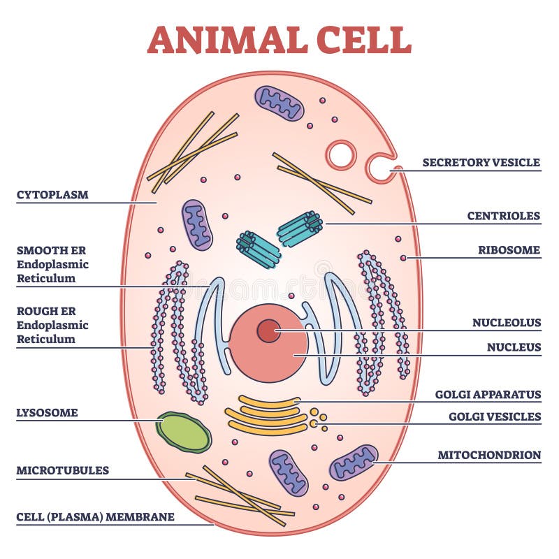
Microfilaments bear a resemblance to microtubules but microfilaments are softer and smaller in diameter. Our body starts its existence at fertilization from a single cell, the diploid zygote. Animal cells are the types of cells that make up most of the tissue cells in animals. Animal cell anatomy label me! Microfilaments are organelle cells formed from actin and myosin proteins. The outer diameter of a microtubule is between 23 and 27 nm while the inner diameter is between 11. Tubulin labeling in live cells with minimal cytotoxicity. Microtubules can grow as long as 50 micrometres and are highly dynamic. Label the animal cell diagram, with a glossary of animal cell terms included. Somewhat like an entire city in miniature. A cell is a complete functional biological unit with many different internal structures. Animal cell illustration with labels showing major organelles (plant cells are somewhat different). Microscopic hollow tubes found inside eukaryotic cells and some prokaryotic bacteria cells that provide structure and motor functions.
Most of the cells size range between 1 illustrated in figure 2 is a pair of fibroblast deer skin cells that have been labeled with fluorescent probes microtubules. Different kinds of animals have microtubules: In animal cells, microtubules arise from centrosomes; Microtubules are exactly how they sound: Our body starts its existence at fertilization from a single cell, the diploid zygote.
A Tour Of The Animal Cell Campbell Biology Diagram Quizlet from o.quizlet.com Also cilia and flagella are made of microtubules. The microtubule cytoskeleton is an essential regulator of the cell cycle, and. Conduit pt et al., centrosome function and assembly in animal cells. Microtubules also form cell structures called centrioles and asters. In animal cells, microtubules radiate outwards from an organelle in the center of the cell called a centrosome, which is a microtubule organizing center (mtoc). Somewhat like an entire city in miniature. Our body starts its existence at fertilization from a single cell, the diploid zygote. Animal cells from the basic structural units of all tissues and organs of the body.
Animal cells are the types of cells that make up most of the tissue cells in animals.
Animal cell anatomy label me! Cell biologist thomas maresca and senior research fellow vikash verma at the university of massachusetts amherst say they have, for the first time, directly observed and recorded in animal cells a pathway called branching microtubule nucleation. The fungi and the majority of plant cells lack centrosomes. The cellular organization of microtubules varies between cell types, but in most cells, the minus ends of microtubules are anchored to the centrosomes near the nucleus while the plus ends radiate towards the periphery of the cell. Animal cells are common names for eukaryotic cells that make up animal tissue. Microtubules are exactly how they sound: Animal cells are the types of cells that make up most of the tissue cells in animals. Start studying animal cell (labeled). One of the primary concerns with using fluorescent taxanes to label microtubules is the possibility of cytotoxicity upon continuous incubation. Microtubules allow motor proteins like kinesin and dynein to carry vesicles (packages of stuff that will be delivered to a different place in the cell). Animal cell microtubules | free images at clker.com. In animal cells, microtubules arise from centrosomes; Microtubules are nucleated and organized by microtubule organizing centers (mtocs), such as the centrosome found in the center of many animal cells or the basal bodies found in cilia and flagella, or the spindle pole bodies found in fungi.
Berbagi :
Posting Komentar
untuk "Animal Cell Labeled Microtubules : Diagram Of Animal Cell Anatomy Stock Vector Illustration Of Reticulum Drawing 120008456 / Most of the cells size range between 1 illustrated in figure 2 is a pair of fibroblast deer skin cells that have been labeled with fluorescent probes microtubules."


Posting Komentar untuk "Animal Cell Labeled Microtubules : Diagram Of Animal Cell Anatomy Stock Vector Illustration Of Reticulum Drawing 120008456 / Most of the cells size range between 1 illustrated in figure 2 is a pair of fibroblast deer skin cells that have been labeled with fluorescent probes microtubules."