Beranda
/ Microtubules In Animal Cell Diagram / The cell. 7. Cytosol. Cytoskeleton. Microtubules. Atlas of ... : Cell what is the function of the microtubules in an animal cell?
Microtubules In Animal Cell Diagram / The cell. 7. Cytosol. Cytoskeleton. Microtubules. Atlas of ... : Cell what is the function of the microtubules in an animal cell?
Microtubules In Animal Cell Diagram / The cell. 7. Cytosol. Cytoskeleton. Microtubules. Atlas of ... : Cell what is the function of the microtubules in an animal cell?. In animal cells, microtubules radiate outwards from an organelle in the center of the cell called a centrosome, which is a microtubule organizing center (mtoc). Most animal cells are diploid, meaning that their chromosomes exist in homologous pairs. Microtubules also form cell structures called centrioles and asters. Microtubules in the cell consist of microscopic structures formed in hollow tubes and constructed in a series of linear rings. Plant cell and animal cell fall under eukaryotic type.
Start studying microtubule organizing centers (mtocs). Chromosomes are attached to kinetochore microtubules via a. Recent findings have shed light on their relative contributions to. We are pleased to provide you with the picture named animal cell diagram in detail. Microtubules are made in the centrosome.
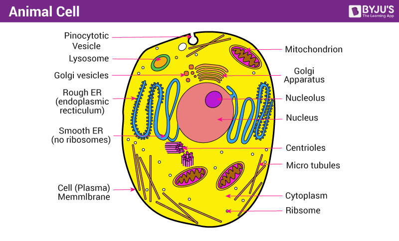
Yoga to your core: Cells, prana and apana from 4.bp.blogspot.com Animal cell illustration with labels showing major organelles including: Lets us discuss the animal cell, types of an animal cell, animal cell diagram, its structure. There are three microtubules in each group. In these cells, microtubules are nucleated from discrete sites in the cytoplasm. You may also find peroxisome, microvilli, microtubules, intermediate filaments, microfilaments, centrosome, flagellum, rough er, smooth er, endoplasmıc reticulum in this image. They are filamentous structures made of soluble. Plant cell and animal cell fall under eukaryotic type. Our body starts its existence at fertilization from a single cell, the diploid zygote.
They help guide the movement the vacuoles in animal cells are generally smaller than that in plant cells.
The walls of the microtubule are made of with a diameter of about 25 nm, microtubules are the widest components of the cytoskeleton. Microtubules as their name implies, microtubules are small hollow tubes. Other cell types, such as trypanosomatid parasites, have a mtoc but it this diagram depicts the organization of a typical mitotic spindle found in animal cells. Each centriole is a ring of nine groups of fused microtubules. A microtubule is found inside some cells but is not a. Animal cells are generally smaller than plant cells and lack a cell wall and chloroplasts; Microtubules are polymers of tubulin that form part of the cytoskeleton and provide structure and shape to eukaryotic cells. Cell what is the function of the microtubules in an animal cell? Most animal cells are diploid, meaning that their chromosomes exist in homologous pairs. The conventional mtoc in animal cells is the centrosome, an organelle next to the nucleus (see chapter 2.5.5). All organisms are made up of cells (or in some cases, a single cell). Start studying microtubule organizing centers (mtocs). Conduit pt et al., centrosome function and assembly in animal cells.
A microtubule is found inside some cells but is not a. Cell biologist thomas maresca and senior research fellow vikash verma at the university of massachusetts amherst say they have, for the first time, directly observed and recorded in animal cells a pathway called branching microtubule nucleation, a mechanism in cell division that had been. The walls of the microtubule are made of with a diameter of about 25 nm, microtubules are the widest components of the cytoskeleton. Animal cells are generally smaller than plant cells and lack a cell wall and chloroplasts; Animal cells are the types of cells that make up most of the tissue cells in animals.
About Microtubule - Assignment Point from www.assignmentpoint.com Our body starts its existence at fertilization from a single cell, the diploid zygote. All organisms are made up of cells (or in some cases, a single cell). These hollow rods help give structure and shape to the cell. In an animal cell, it is since eukaryotic cells greatly depend upon the integrity of microtubules and other cytoskeletal filaments to. As observed in the labeled animal cell diagram, the cell membrane forms the confining factor of the cell, that is it envelopes the cell constituents together and gives the cell its shape, form, and existence. The cell is the basic unit of life. A microtubule is found inside some cells but is not a. These are organelles pertinent to microtubules are straight hollow filaments that act like support beams.
Chromosomes are attached to kinetochore microtubules via a.
Animal cells are generally smaller than plant cells and lack a cell wall and chloroplasts; Microtubules are polymers of tubulin that form part of the cytoskeleton and provide structure and shape to eukaryotic cells. Microtubules are made in the centrosome. Our body starts its existence at fertilization from a single cell, the diploid zygote. In the complete animal cell centrosome, the two centrioles are arranged such that one is perpendicular to. You may also find peroxisome, microvilli, microtubules, intermediate filaments, microfilaments, centrosome, flagellum, rough er, smooth er, endoplasmıc reticulum in this image. Microtubules can also be distinguished from microfilaments chemically. They help guide the movement the vacuoles in animal cells are generally smaller than that in plant cells. The cell is the basic unit of life. The diagram, like the one above, will include labels of the major parts of an animal cell including the cell membrane, nucleus, ribosomes, mitochondria, vesicles, and cytosol. Lets us discuss the animal cell, types of an animal cell, animal cell diagram, its structure. Microtubules in the cell consist of microscopic structures formed in hollow tubes and constructed in a series of linear rings. The cellular organization of microtubules varies between cell types, but in most cells, the minus ends of microtubules are anchored to the centrosomes near the nucleus while the plus ends radiate towards the periphery of the cell.
Microtubules are made in the centrosome. Cell membrane is made up of lipids and proteins and forms a barrier between the extracellular liquid. These are organelles pertinent to microtubules are straight hollow filaments that act like support beams. A microtubule is found inside some cells but is not a. The outer diameter of a microtubule is between 23 and 27 nm while the inner diameter is between 11.
Animal Cell - Structure, Function and Types of Animal Cell from cdn1.byjus.com Microtubules (and centrioles) are part of the cytoskeleton. Microtubules are present in both plant cells and animal cells and are included in courses in cell biology. Microtubules as their name implies, microtubules are small hollow tubes. In animal cells, microtubules radiate outwards from an organelle in the center of the cell called a centrosome, which is a microtubule organizing center (mtoc). You may also find peroxisome, microvilli, microtubules, intermediate filaments, microfilaments, centrosome, flagellum, rough er, smooth er, endoplasmıc reticulum in this image. Lets us discuss the animal cell, types of an animal cell, animal cell diagram, its structure. Microtubules are made in the centrosome. The cell is the basic unit of life.
Microtubules are found in the cytoplasm of all types of eukaryotic cells with rare absence, such as in human erythrocytes.
The cell is the basic unit of life. Microtubules are present in all eukaryotic cells and have been found to play a variety of structural and dynamic (c) diagram of the role of microtubule motors in cgn migration. Microtubules also form cell structures called centrioles and asters. In these cells, microtubules are nucleated from discrete sites in the cytoplasm. There are three microtubules in each group. Start studying microtubule organizing centers (mtocs). Our body starts its existence at fertilization from a single cell, the diploid zygote. Both of these structures are found in animal cells, but not plant cells. Cell membrane is made up of lipids and proteins and forms a barrier between the extracellular liquid. Cell biologist thomas maresca and senior research fellow vikash verma at the university of massachusetts amherst say they have, for the first time, directly observed and recorded in animal cells a pathway called branching microtubule nucleation, a mechanism in cell division that had been. The outer diameter of a microtubule is between 23 and 27 nm while the inner diameter is between 11. Microtubules as their name implies, microtubules are small hollow tubes. Microtubule length is quite variable.
Berbagi :
Posting Komentar
untuk "Microtubules In Animal Cell Diagram / The cell. 7. Cytosol. Cytoskeleton. Microtubules. Atlas of ... : Cell what is the function of the microtubules in an animal cell?"


Posting Komentar untuk "Microtubules In Animal Cell Diagram / The cell. 7. Cytosol. Cytoskeleton. Microtubules. Atlas of ... : Cell what is the function of the microtubules in an animal cell?"