Typical Animal Cell Under Light Microscope : enbrocunlex: Animal Cell Under Light Microscope - Typical animal cell pinocytotic vesicle lysosome golgi vesicles golgi vesicles rough er (endoplasmic reticulum) smooth er (no ribosomes) cell (plasma) membrane mitochondrion golgi apparatus nucleolus nucleus centrioles (2) each composed of 9 microtubule triplets microtubules.. We use microscope comprehensively in microbiology, mineralogy, cell biology, biotechnology, nano physics, microelectronics, pharmacology, and forensics. Lymphocytes are the cells which are comparatively smaller in size and under the microscope appear spherical in shape with. Typical plant cell (nucleus and membrane… Typical animal cell pinocytotic vesicle lysosome golgi vesicles golgi vesicles rough er (endoplasmic reticulum) smooth er (no ribosomes) cell (plasma) membrane mitochondrion golgi apparatus nucleolus nucleus centrioles (2) each composed of 9 microtubule triplets microtubules. Most cells are very small, and their structures can only be seen by using a microscope.
What can only be seen under a microscope can now cover an entire serving plate. As for seeing electrons under any microscope in general, i would say we have come as close to it as scientifically and technically possible with the tem having a resolution of 2 nm (there plant cells look pretty much like animal cells except they have a cell wall and chloroplasts for photosynthesizing. Typical plant cell (nucleus and membrane… Under the microscope, an animal cell shows many different parts called organelles, that work together to keep the cell functional. Observe the onion skin under low power of the microscope and then under high power.
How & Why Fasting Can Boost Your Immune System « Food ... from img.wonderhowto.com Eukaryotic cells make up the tissues of all plants and animals. Light rays pass through the specimen on a slide. Microscopes can be simple or complex in design, and some can do more than one type of microscopy, each of which reveals slightly different information. smaller dots within the cytoplasm are particles of stored food. 15 видео 74 483 просмотра обновлен 16 апр. Cells consist of cytoplasm enclosed within a membrane, which contains many biomolecules such as proteins and nucleic acids.2 most plant and animal cells are only visible under a light microscope, with dimensions between 1 and 100 micrometres.3 electron microscopy gives a much higher. A cell is a very tiny structure which exists in living bodies. Examining animal cells under the microscope.
Observing a wide range of biological processes and animal cell under light microscope is easier due to advances in microscopic techniques.
You have recently studied a cartoon that illustrates the basic structure and organelles of a typical animal cell. Animal cell cake of celliness: Focused by an objective lens and an eyepiece lens. Typical plant cell (nucleus and membrane… As for seeing electrons under any microscope in general, i would say we have come as close to it as scientifically and technically possible with the tem having a resolution of 2 nm (there plant cells look pretty much like animal cells except they have a cell wall and chloroplasts for photosynthesizing. Lymphocytes are the cells which are comparatively smaller in size and under the microscope appear spherical in shape with. The parts of a (palisade) plant cell that can be seen under a light microscope are:cell wallcell (surface) membranelarge (permanent) vacuolecytoplasmnucleuschloroplasts. The light microscope also provides a better view of the mouthparts of the ant. What can u observed under the light microscope. When we view a specimen under a microscope it needs to let light pass through the specimen so we can see it. Learn how to make an animal cell cake! All living things are composed of cells. Under the microscope, animal cells appear different based on the type of the cell.
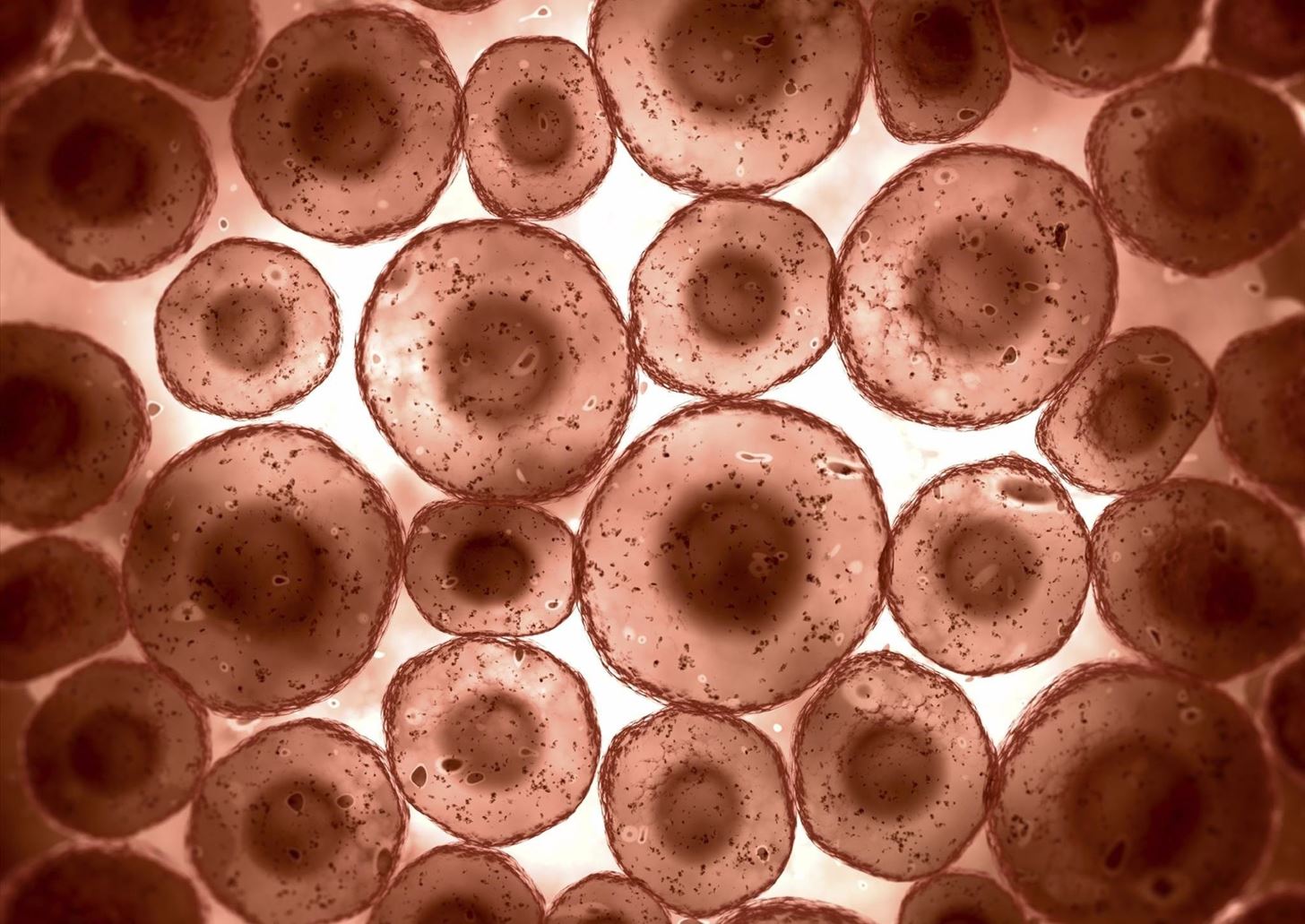
Typical animal cell (membranes) light microscope. Light uses light waves as it's source of radiation and electron microscopes use electrons. With a light microscope you can see several structures inside the cell. Learn how to make an animal cell cake! Microscopes can be simple or complex in design, and some can do more than one type of microscopy, each of which reveals slightly different information.
Slide, Microscope, Animal Cell from shop.omegascientific.com.au Light and electron microscopes allow us to see inside cells. All living things are composed of cells. Light rays pass through the specimen on a slide. Eukaryotic cells make up the tissues of all plants and animals. An iris diaphragm on the substage condenser controls the amount of light reaching the objective, and also affects the contrast of the specimen. 15 видео 74 483 просмотра обновлен 16 апр. Light uses light waves as it's source of radiation and electron microscopes use electrons. Lymphocytes are the cells which are comparatively smaller in size and under the microscope appear spherical in shape with.
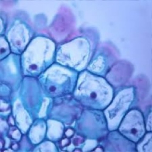
Lymphocytes are the cells which are comparatively smaller in size and under the microscope appear spherical in shape with.
The eyepiece and an objective lens. This diagram shows a typical animal cell. Cells consist of cytoplasm enclosed within a membrane, which contains many biomolecules such as proteins and nucleic acids.2 most plant and animal cells are only visible under a light microscope, with dimensions between 1 and 100 micrometres.3 electron microscopy gives a much higher. Cell structure teaching resources the science teacher, organelles biology for majors i, 11 different types of cells in the human body, class test, chronic inflammation under the microscope learn share. Light rays pass through the specimen on a slide. We use microscope comprehensively in microbiology, mineralogy, cell biology, biotechnology, nano physics, microelectronics, pharmacology, and forensics. All living things are composed of cells. The parts of a (palisade) plant cell that can be seen under a light microscope are:cell wallcell (surface) membranelarge (permanent) vacuolecytoplasmnucleuschloroplasts. Are you more like an animal cell or a plant cell? Plant, animal and bacterial cells have smaller components calculating the magnification of light microscopes. Lymphocytes are the cells which are comparatively smaller in size and under the microscope appear spherical in shape with. Plant cells have a fixed shape. Learn how to make an animal cell cake!
15 видео 74 483 просмотра обновлен 16 апр. Gcse biology microscope drawing and measuring cell size edexcel 9 1. Observe the onion skin under low power of the microscope and then under high power. We use microscope comprehensively in microbiology, mineralogy, cell biology, biotechnology, nano physics, microelectronics, pharmacology, and forensics. A cell is a very tiny structure which exists in living bodies.
BIOLOGY ORDINARY LEVEL NOTES: CELL STRUCTURES from 1.bp.blogspot.com Focused by an objective lens and an eyepiece lens. Plant, animal and bacterial cells have smaller components calculating the magnification of light microscopes. Learn how to make an animal cell cake! An iris diaphragm on the substage condenser controls the amount of light reaching the objective, and also affects the contrast of the specimen. What can u observed under the light microscope. Under the microscope, an animal cell shows many different parts called organelles, that work together to keep the cell functional. Lymphocytes are the cells which are comparatively smaller in size and under the microscope appear spherical in shape with. A cell is a very tiny structure which exists in living bodies.
What was once unseeable can now be seen, touched, and eaten!cut yourself a wedge for dessert or snack on a nucleus, lyosome, or…
As for seeing electrons under any microscope in general, i would say we have come as close to it as scientifically and technically possible with the tem having a resolution of 2 nm (there plant cells look pretty much like animal cells except they have a cell wall and chloroplasts for photosynthesizing. Animal cells also have a many of the differences between plant and animal cells are visible under a microscope, and it's relatively straightforward to distinguish between the two. Examining animal cells under the microscope. Observe the onion skin under low power of the microscope and then under high power. Plant cells have a fixed shape. This diagram shows a typical animal cell. To prepare and study a slide of animal cells (human. Light and electron microscopes allow us to see inside cells. Plant cells have cell walls, one large vacuole per cell, and chloroplasts, while animal cells will have a cell membrane only. When we view a specimen under a microscope it needs to let light pass through the specimen so we can see it. Microscopes can be simple or complex in design, and some can do more than one type of microscopy, each of which reveals slightly different information. (2) study of animal cells : An animation that shows animal cells.
Berbagi :
Posting Komentar
untuk "Typical Animal Cell Under Light Microscope : enbrocunlex: Animal Cell Under Light Microscope - Typical animal cell pinocytotic vesicle lysosome golgi vesicles golgi vesicles rough er (endoplasmic reticulum) smooth er (no ribosomes) cell (plasma) membrane mitochondrion golgi apparatus nucleolus nucleus centrioles (2) each composed of 9 microtubule triplets microtubules."


Posting Komentar untuk "Typical Animal Cell Under Light Microscope : enbrocunlex: Animal Cell Under Light Microscope - Typical animal cell pinocytotic vesicle lysosome golgi vesicles golgi vesicles rough er (endoplasmic reticulum) smooth er (no ribosomes) cell (plasma) membrane mitochondrion golgi apparatus nucleolus nucleus centrioles (2) each composed of 9 microtubule triplets microtubules."